Foreword:
Lasers are an increasingly important tool in modern bioimaging and analysis systems, including flow cytometry to confocal and multiphoton microscopy. However, in many commercial imaging systems, performance is severely limited by defects such as laser wavelength, cost, power, and reliability inherent in conventional laser technology. The ultra-fast fiber laser provided by Shenzhen Optical Laser provides you with a "turnkey", ultra-small, high-reliability solution that is the best source for next-generation biomedical imaging systems.
Ultra-fast fiber lasers are characterized by ultra-small form factor, inherent high reliability and low maintenance cost, and are increasingly popular laser sources for many biomedical imaging applications. Traditional titanium-sapphire lasers present serious challenges and create significant competitive pressures for conventional semiconductor, helium, and argon lasers in the field of fluorescence analysis.
The ultra-fast fiber laser of Shenzhen Optical Domain Laser adopts MOPA structure, which greatly simplifies and flexes the design requirements. The MOPA structure consists of two separate components: a low-power femtosecond or picosecond fiber laser (MO) and a high-power fiber amplifier (PA). A laser with different optical parameters and performance can be obtained independently by changing the parameters of the host vibration laser (MO) or amplifier (PA) to meet your specific and specified application requirements. This flexible design structure has enabled ultrafast fiber lasers to quickly meet a wide range of application needs across a wide range of applications.
Typical characteristics of ultrafast fiber lasers:
The main vibrator (MO) is typically an all-fiber passive mode-locked laser that requires no adjustments and can be operated with a key switch and self-start. Pulse frequencies (<1Mhz to hundreds of Mhz) are ideal for quasi-continuous laser applications and for life imaging applications.
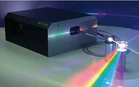
Figure 1: Partial spectrum of visible light emitted by a diffraction grating
In many applications of microscopy, ultrafast lasers often involve multiphoton fluorescence microscopy. In these applications, Ti:Sapphire lasers are a natural choice due to the history of laser development. However, we all know that Ti:Sapphire lasers cannot provide a tunable broadband spectrum, but the femtosecond fiber lasers of Shenzhen Optical Domain Lasers can provide discrete wavelengths in the 980nm-1100nm range. They also have the advantages of low cost and small size. The potential to be compatible with your existing microscopy system.
Today, "turnkey" lasers with an average output power of up to 5W and pulse widths shorter than 250fs are already available. The fiber-optic structure of the laser system has a super long wavelength range that exceeds the tunable limit of most titanium sapphire lasers, bringing many benefits to two-photon fluorescence and second harmonic microscopy.
High-power fiber lasers operating in picosecond or femtosecond states, with single-pulse energy up to pico-joules to tens of micro-joules. This parameter is especially important for material processing and is equally important for component parts and biological tissue.
The nonlinear frequency conversion technology can broaden the radiation spectrum of the fiber laser from near-infrared to visible to ultraviolet, and obtain 532nm, 355nm and 266nm wavelength output, and can also produce a super-wide "supercontinuum spectrum"----this phenomenon will There will be a huge “brainwashing shock†for the next generation of biomedical imaging systems.
Supercontinuum laser----the latest development of laser history
The generation of supercontinuum spectra in highly nonlinear optical fibers such as photonic crystal fibers or tapered fibers was discovered by Lucent and the University of Bath in the United Kingdom in 1999-2000, when the continuous spectrum was demonstrated from 400 nm to 2000 nm. Early fiber optic supercontinuum sources were achieved by freely spatially coupling high-intensity femtosecond pulses (such as Ti:Sapphire lasers) or nanosecond-order pulsed light (such as Q-switched chip lasers) into photonic crystal fibers. However, when it is necessary to provide a true white laser for the laboratory, it is found that the laser with the above high-intensity light for end-coupling into a very fine fiber (3um in diameter) has many unstable phenomena. And congenital problems such as unreliability, so that the process technology can not meet the actual requirements of real commercialization.
In 2005, the British cooperation company of Shenzhen Optical Laser developed a mature supercontinuum fiber laser. At that time, its spectrum covered 450nm to 2000nm, and the average power was close to 2W. The supercontinuum fiber laser is based on a high power picosecond source and a length of highly nonlinear photonic crystal fiber. The high-density pulsed light is tightly constrained in a narrow-spaced optical fiber waveguide, resulting in a supercontinuum spectrum from visible light (below 400 nm) to 2000 um. Based on the all-fiber structure, the laser system is coupled by a mature and simple fiber fusion, rather than the free-space end face projection into the fiber in the traditional sense, thus greatly improving its reliability and stability. The problem is the difficulty that almost other fixed lasers can't overcome when producing supercontinuum spectra.
As a product that has been commercialized for 4 years, the super-continuous fiber laser of Shenzhen Optical Domain has been improved and improved in many aspects. In 2005, our new laser system broke the limits on wavelength and output power. Table 1 clearly compares the laser parameters of ours today and 2005. For the past 3 years we have focused on improving laser brightness and shorter wavelength output, as these two parameters are critical and fundamental requirements in many imaging applications. The latest (2007) improvement is to increase the spectral density to several mW/nm in the full range from 400 nm to 2000 nm (see Figure 1). However, perhaps the most important improvement is the reliability of the laser, which is satisfactorily certified by our many field demonstration applications, from fluorescence imaging to nano-optics to basic research.
This paper focuses on the latter half of the key biomedical technology used in these applications of our fiber supercontinuum laser such as: blood cells flow technology, and confocal microscopy optical coherence tomography.
A laser solves the fluorescence into a rubber
Flow cytometry (FACS) is a key tool for biomedical research. These complex instruments are used to measure the properties of individual cells, typically by detecting fluorescent molecules (fluorophores) attached to their surface. These fluorescent molecules are like a probe that plays a crucial role in the analysis of the cellular immune system, the identification of cancer cells and the diagnosis of diseases. The progress of flow cytometry relies almost entirely on the laser beam to excite the fluorophore. However, the coherence and power levels of these lasers make them ideal for light-emitting single cells, but their discontinuous light waves limit the types of fluorescent probes that can be analyzed by flow cytometry.
Although solid-state laser technology has increased the diversity of discontinuous wavelength lasers, there is still a huge gap between the range of light waves that they can accept with fluorescence excitation, which greatly limits the ability to perform fluorescence detection in biomedical analysis.
Confocal fluorescence microscopy is one of the most powerful techniques for detecting a wider range of complex medical phenomena in the biomedical sciences, where laser sources such as flow cytometry are also required.
Almost all current commercial confocal microscopy systems are fluorescently excited by combining several discrete wavelength lasers in the vicinity of visible light. However, in flow cytometry, the excitation wavelength is actually a small part by using a laser with a fixed wavelength (even several). Many fluorophores cannot be detected and utilized due to this limitation.
In order to promote the development of fluorescence excitation technology, multi-channel AOTF technology (acoustic-light modulation filter) and super-continuous fiber laser have been developed in recent years. A supercontinuum fiber laser with an 8-channel AOTF provides eight laser beams in the visible range, each of which can be modulated over the entire spectral range and is in the same line at the same divergence diffraction limit. on. The combination of supercontinuum spectroscopy and AOTF technology provides a very flexible solution for flow cytometry or fluorescence confocal microscopy for the optimization of a wide range of excitation and detection. The wavelength of this laser can be analyzed by precision tuning to the excitation peak of the fluorescent probe.
What's more, the supercontinuum fiber laser provided by Shenzhen Optical Field Laser not only has a significant improvement in performance, but also has a huge advantage in cost. For example, on the flow cytometry experimental platform shown in Figure 2, except for a triple-frequency Nd:YVO4 355nm UV laser, all other lasers and many optics can use only one supercontinuum fiber laser. replace. At the same time, with the continuous development of supercontinuum fiber laser technology, it is only a matter of time before the spectral range extends to the ultraviolet band. At that time, a supercontinuum fiber laser can replace all lasers, and even meet the highest specification imaging system. A variety of continuous spectra are required.
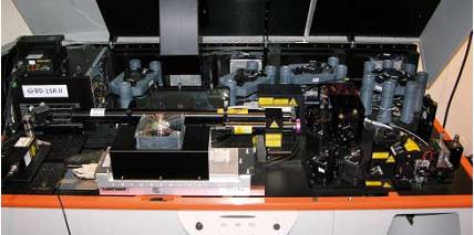
Figure 2: A high-standard flow cytometry table consisting of eight lasers with discontinuous wavelengths can be replaced by a supercontinuum laser.
However, commercial units engaged in confocal microscopy technology will soon integrate supercontinuum fiber lasers into their imaging systems, and they are generally the first research institutes to invest in this advanced technology. For example, Dr. Clemens Kaminski of the Laser Analysis Group in Cambridge, England, and his team recently integrated a supercontinuum fiber laser for 2D and 3D and live cell imaging in his Olympus FluoView microscope scanning system. the study.
Figure 3 shows a sample of two-dimensional high-precision fluorescence confocal imaging using SC450 series supercontinuum laser and AOTF system using Shenzhen optical domain laser. In this experiment, a lily of the valley specimen was stained with red and green, with peak excitation wavelengths of 530 nm and 620 nm, respectively. Since the two dyes have many affinity relationships with other regions of the sample, scanning the excitation wavelength can highlight different regions of the sample. Fibrous cell walls appear throughout the sample after staining with green (both 620 nm and 640 nm images), while lignin stained with saffron dye appears in the endothelium and xylem, and is particularly visible in the excitation range from 540 nm to 560 nm.
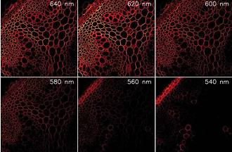
Figure 3: Separate imaging of the rhizome of lily of the valley flowers after blush and green fixation at 530 nm and 620 nm peak excitation bands
Optical coherence tomography
Optical coherence tomography (OCT) is an interferometric and non-invasive imaging technique that provides infiltration of millimeters (up to 3 mm in biological tissue) with longitudinal accuracy typically on the order of micrometers. The application of supercontinuum sources in OCT is another important application in the field of biomedical imaging.
The most common practice in traditional OCT systems is to use a super-cold-light semiconductor laser as the light source, but it is severely limited by the bandwidth, so the longitudinal accuracy of imaging is generally only 10-15 microns. Ultra-precise OCT systems can be implemented with ultra-expensive but super-complex femtosecond solid-state lasers with sub-micron longitudinal accuracy at the 725 nm band.
Shenzhen's optical domain laser's "turnkey" supercontinuum fiber lasers enable higher spectral bandwidths and ever-increasing output power while maintaining constant reliability.
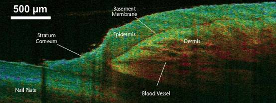
Felix Spoler of the Institute of Semiconductor Electronics in Aachen, Germany, and his colleagues have recently used the high brightness and wide bandwidth of super-connected fiber lasers to demonstrate ultra-high-precision OCT systems. Their approach is to split the supercontinuum fiber laser into two bandwidths at the center of the 840 nm and 1230 nm, each with an average power of more than 100 mW, and each band has a bandwidth of nearly 200 nm. The 840nm and 1230nm dual-wavelength OCT systems take full advantage of the high longitudinal accuracy of short-wavelength illumination and high penetration depth at long wavelengths. The implementation of this technique can achieve 1.8um accuracy (840nm) and 3.8um precision (1230nm) in free space, respectively. In addition, the simultaneous use of two wavelengths also causes mixing at each wavelength OCT imaging, thereby reducing imaging spots. Imaging with different spectra can also increase the contrast of the image, so each wavelength band can be analyzed by the backscattered light of the sample, providing another standard of information for the structure of the sample.
Figure 4 shows the different spectral imaging of a human nail in vivo, showing different levels and blood vessels in the modified area. Here, the difference in the scattering intensity of the two wavelength bandwidths is marked by color.
Outlook
Biomedical applications such as flow cytometry, optical coherence tomography, CARS microscopy, and confocal fluorescence microscopy have enormous practical demands for suitable broad-spectrum illumination sources, which continue to drive the development of laser technology.
In the past five years, ultrafast fiber lasers have begun to challenge traditional solid-state lasers, semiconductor lasers and gas lasers in many application markets, and it is expected that as laser technology continues to evolve and costs decrease, as the next generation of biomedical The best light source for the rubber system, supercontinuum fiber laser will be more and more widely promoted and applied. (wenjing)
Chunmee 4011 is one of the treasures of Green Tea.The shape is tightly tied, well-balanced, grey-green frost, oily and fragrant. The picking standard of fresh leaves is the initial development of one bud, one leaf and one bud and two leaves. For those with thick white hair and strong buds, six processes are selected, such as spreading, killing, rolling, baking, frying and re-drying. Chunmee 4011 Green Tea has special quality and health function, its effect is like rhinoceros horn, has cool and refreshing detoxification, bright eye decrease internal heat's wonderful effect, can treat "fire disease".
Chunmee 4011 are produced by Joy Tea Co.,LTD with high quality and good appearance. Welcome you to visit our company. For inqury, please send mail directly.

Chunmee 4011 Green Tea
Chunmee 4011 Green Tea,Organic Chunmee Green Tea 4011,Health Chunmee Green Tea 4011,Non-Polluted Chunmee Green Tea
JOY TEA CO., LTD. , https://www.joyteaco.com